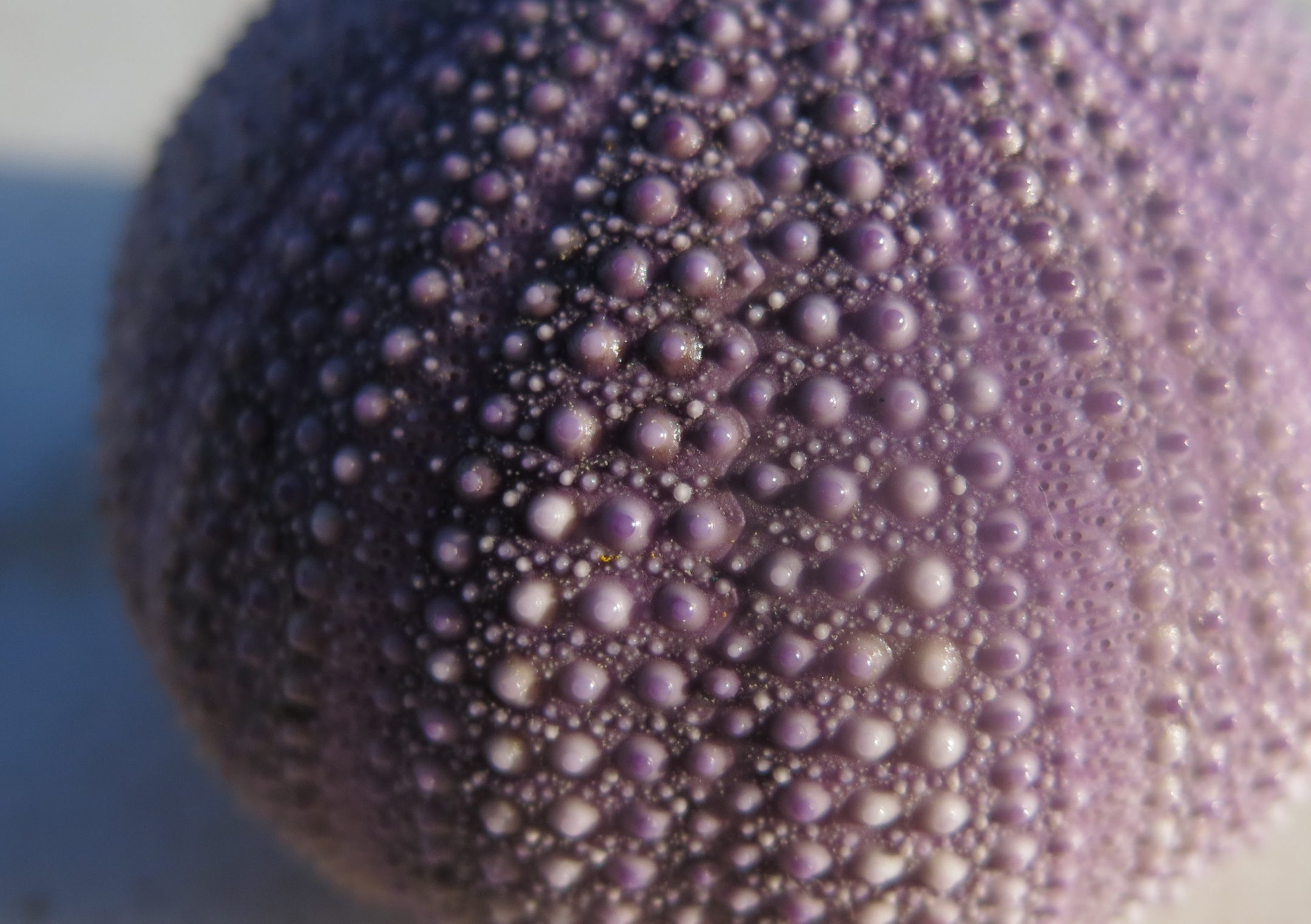

The calcite crystals in purple sea urchin teeth are co-oriented thanks to propagation of existing crystallinity through an amorphous precursor.
“Sea urchin teeth are remarkable and complex calcite structures,
continuously growing at the forming end and self-sharpening at the
mature grinding tip. The calcite (CaCO3) crystals of tooth
components, plates, fibers, and a high-Mg polycrystalline matrix, have
highly co-oriented crystallographic axes. This ability to co-orient
calcite in a mineralized structure is shared by all echinoderms.
However, the physico-chemical mechanism by which calcite crystals become
co-oriented in echinoderms remains enigmatic. Here, we show differences
in calcite c-axis orientations in the tooth of the purple sea
urchin (Strongylocentrotus purpuratus), using high-resolution
X-ray photoelectron emission spectromicroscopy (X-PEEM) and microbeam
X-ray diffraction (μXRD). All plates share one crystal orientation,
propagated through pillar bridges, while fibers and polycrystalline
matrix share another orientation. Furthermore, in the forming end of the
tooth, we observe that CaCO3 is present as amorphous calcium
carbonate (ACC). We demonstrate that co-orientation of the
nanoparticles in the polycrystalline matrix occurs via solid-state
secondary nucleation, propagating out from the previously formed fibers
and plates, into the amorphous precursor nanoparticles. Because
amorphous precursors were observed in diverse biominerals, solid-state
secondary nucleation is likely to be a general mechanism for the
co-orientation of biomineral components in organisms from different
phyla.” (Killian et al. 2009:18404)
http://www.news.wisc.edu/17493
http://pubs.acs.org/doi/abs/10.1021/ja907063z