


Nematocysts of some cnidarians can penetrate thick layers of crustacean shell by capsules of unusually short collagens that explosively eject stylets of strong and flexible protein tubules with spiked barbs.
Introduction
Overcoming the protective cuticle of armored opponents is a challenge faced by many organisms for both defensive and predatory reasons. Thick shells like those of crustaceans are especially difficult to penetrate without the aid of sharp and strong body parts and powerful muscles to back them up. Some organisms solve this challenge with only microscopic cellular components. Hydras, tiny animals from the cnidarian genus Hydra, feed on planktonic crustaceans and have evolved remarkable nano-structures that can penetrate the armor of their prey to inject venom.
The Strategy
The cells (cnidocysts) produce one large organelle called a nematocyst. The cell forms a layered matrix around the nematocyst that keeps it strong and promotes the generation of 150 bar of pressure within the organelle at maturation. The “lid” of the capsule (operculum) associates with the cell membrane facing out. When the sensory portion of the cell (cnidocil) is mechanically disturbed (e.g., by contact with prey) it causes a rapid increase in the calcium ion concentration in the cell. This causes molecular rearrangement of the opeculum allowing the release of the nematocyst’s stored pressure towards the outside of the organism. The stylet, composed of strong and flexible tubules with spiked barbs at the end, ejects from the cell with an acceleration of ~5.4 million times gravity. Because of the tiny cross-sectional area at the tip of the stylet, 7.7 billion Pascals of pressure are exerted upon the cuticle of the prey and drives it deep into the underlying tissue. In fact, this is not only the fastest animal system observed to date, the pressure on impact is also comparable to that produced by a bullet.

A retracted live green hydra (Phylum Cnidaria, class Hydrozoa, genus Hydra) tentacle (cnida) showing multiple nematocysts that are still embedded in the cnidocytes that formed them. Nematocysts of this species have a bulb-shaped base that connects to a narrow barbed top, from which a long thin filament extends many times the length of the nematocysts base. In this image the nematocysts are all still embedded in the cnidocytes, clear cells lining the tentacle that produce nematocysts. Two nematocysts are focused on in this image. Seen at approximately 1,000x magnification under a light microscope with no staining or artificial coloring.

Nematocyst Animation
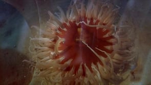
This video animation from Shape of Life depicts the function of nematocysts in the tentacles of an anemone (another type of cnidarian).


